


The space between neurons: a territory to explore to understand brain aging and diseases such as Parkinson's
• July 22 marks World Brain Day
• “Traditionally, research has focused on the sending and receiving neurons. Our approach is to understand how the environment between neurons itself modulates that signal,” says researcher Jan Tønnesen
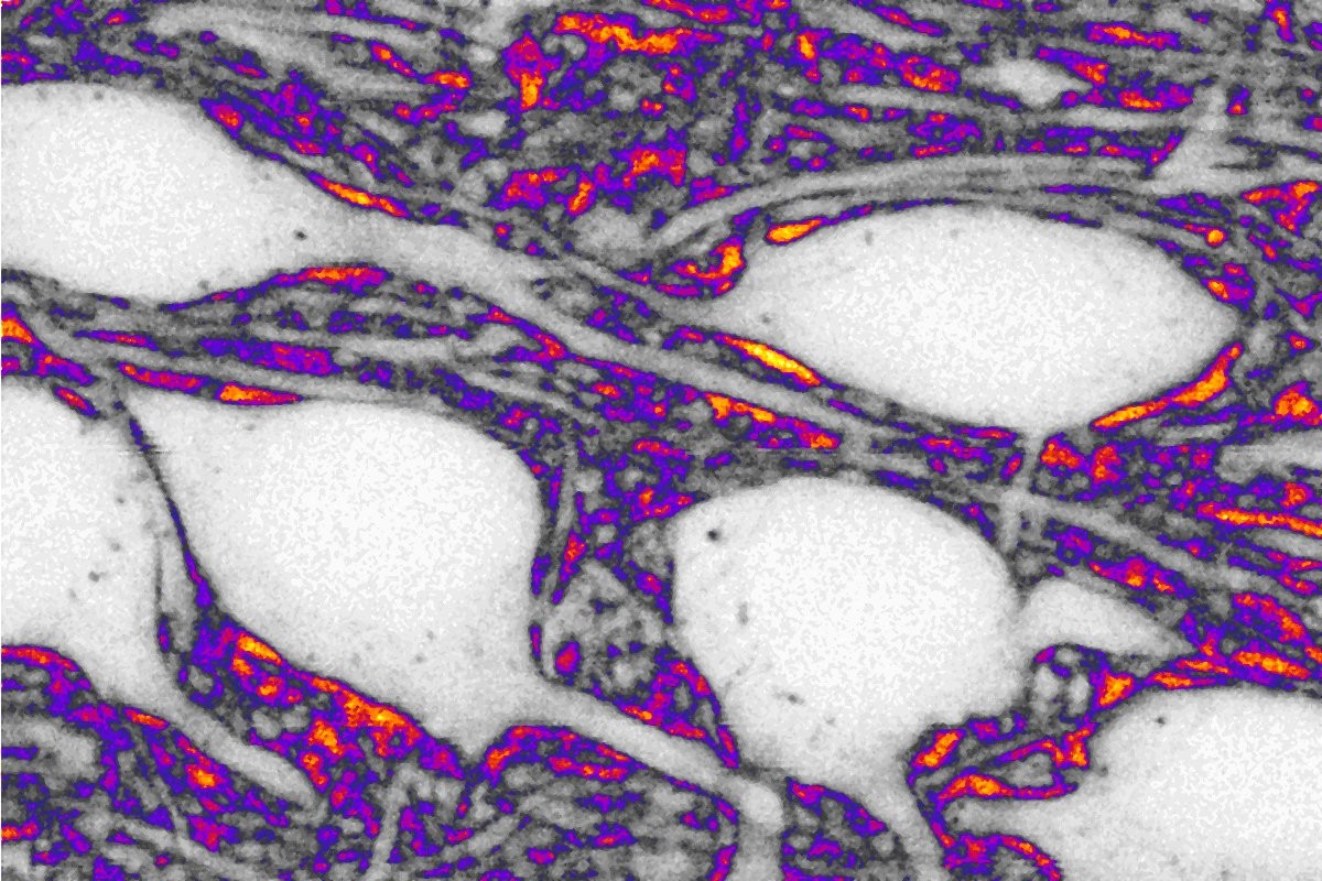
Coinciding with World Brain Day, celebrated on July 22, the Instituto Biofisika (CSIC, EHU) highlights the advances made by one of its teams in understanding the microscopic architecture of the brain. The group led by Jan Tønnesen has succeeded in directly visualizing the extracellular space—the tiny region separating neurons—an area that until now has been virtually inaccessible for live observation.
This space, which is key to neuronal communication, is where neurotransmitters diffuse, enabling the exchange of signals between cells. “Traditionally, research has focused on the sending and receiving neurons. Our approach is to understand how the environment between the neurons itself modulates that signal,” explains Tønnesen, the principal investigator.
This breakthrough has been made possible by a new generation of fluorescence microscopy that allows for the observation of structures at the nanoscale. The technique, known as super-resolution shadow imaging (SUSI), provides negative contrast that reveals the geometry of the extracellular space with great precision. This approach opens the door to studying how its organization influences processes such as cognition, brain aging, and the development of diseases.
“If we are successful, in the future we will be able to better target therapies for neurodegenerative diseases such as Parkinson’s, or understand how age-related and biologically driven deterioration occurs,” says Alejandro Fierro, a predoctoral research fellow in the group. His daily work includes maintaining live cell cultures using specialized media, which allow tissues to be observed under controlled conditions for hours.
The laboratory combines these cultures with solutions designed to replicate the brain’s energy environment, which is highly dependent on glucose. This experimental control facilitates the study of the structure and function of brain tissue under conditions close to physiological ones.
The ability to analyze the brain’s structural complexity at this level of detail represents a significant leap forward. “We can now observe aspects that were previously invisible and better understand how neurons connect and how brain activity is generated,” adds Fierro.
According to the World Health Organization, neurological diseases are one of the leading causes of disability worldwide, affecting hundreds of millions of people. Specifically, Parkinson’s disease affects more than 8.5 million people globally (WHO, 2022). These figures underscore the importance of research aimed at advancing our understanding and treatment of these diseases.