Next BBDG Seminar: "Nanopipette robotics for multiparametric imaging and manipulation of living cells"
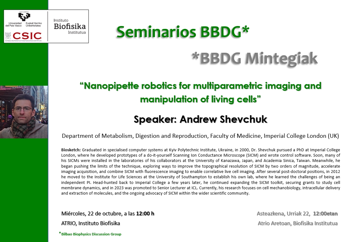
🗓️ October, Wed 22, 2025, 12:00h
📍Atrium (Instituto Biofisika)
📄 "Nanopipette robotics for multiparametric imaging and manipulation of living cells"
🗣️ Andrew Shevchuk (Department of Metabolism, Digestion and Reproduction, Faculty of Medicine, Imperial College London, UK)
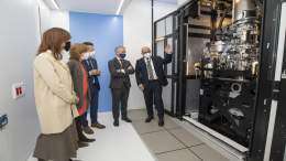
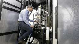
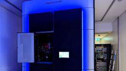
In this seminar, Dr. Andrew Shevchuk will present a decade of advancements in Scanning Ion Conductance Microscopy (SICM) instrumentation and methodologies that have transformed this glass nanopipette scanning technique. Originally used solely to generate three-dimensional topographical images of living cells in physiological buffer media, it has quickly evolved into a multiparametric live imaging tool with active manipulation capabilities. By combining SICM with fluorescence confocal microscopy and, more recently, fluorescence light sheet imaging, Dr. Shevchuk will demonstrate how it is possible to link dynamic volumetric changes in cell 3D nanomorphology with fluorescence signals from specific molecules involved. Furthermore, glass nanopipettes can be chemically modified to become electrochemical nanoprobes for localised measurement of pH, oxygen, ROS, and ATP, many of which can be carried out in a correlative imaging manner. Despite being a truly non-invasive imaging technique capable of, but not limited to, 24-hour live cell topographical imaging, a methodology has been developed to use SICM pipettes to apply forces in the hundreds of piconewtons range for low-stress mapping of cell mechanical properties with nanoscale resolution. SICM nanopipettes can also be used for multiple rounds of controlled delivery and extraction of charged and neutral molecules via localised electroporation into and from the cell cytoplasm and nucleus, with minimal damage, along with detecting DNA, RNA, and proteins through translocation. Coupled with the ability for highly localised drug delivery via nanopipette, dose-response measurements, and electrophysiological recordings, all these features combine to form a versatile, robotic, multitool system suitable for a wide range of experiments aimed at revealing subtle aspects of cell (patho)physiology and/or cell biophysics.
Dr. Shevchuk Biosketch
Graduated in specialised computer systems at Kyiv Polytechnic Institute, Ukraine, in 2000, Dr. Shevchuk pursued a PhD at Imperial College London, where he developed prototypes of a do-it-yourself Scanning Ion Conductance Microscope (SICM) and wrote control software. Soon, many of his SICMs were installed in the laboratories of his collaborators at the University of Kanazawa, Japan, and Academia Sinica, Taiwan. Meanwhile, he began pushing the limits of the technique, exploring ways to improve the topographical resolution of SICM by two orders of magnitude, accelerate imaging acquisition, and combine SICM with fluorescence imaging to enable correlative live cell imaging. After several post-doctoral positions, in 2012 he moved to the Institute for Life Sciences at the University of Southampton to establish his own lab, where he learned the challenges of being an independent PI. Head-hunted back to Imperial College a few years later, he continued expanding the SICM toolkit, securing grants to study cell membrane dynamics, and in 2023 was promoted to Senior Lecturer at ICL. Currently, his research focuses on cell mechanobiology, intracellular delivery and extraction of molecules, and the ongoing advocacy of SICM within the wider scientific community.